
Atomic resolution in situ atomic force microscopy (AFM) images reveal mica lattice (a). (b) shows peptoid fibers. Bottom images are ball-stick model of mica lattice, and Fourier transform of (b).
Tuning nucleation pathways through sequence-engineering of biomimetic polymers
Ma, X., Newcomb, C., Chen, C., & De Yoreo, J.
0000
(2016)
Ma, X., Newcomb, C., Chen, C., & De Yoreo, J.
in prep
00
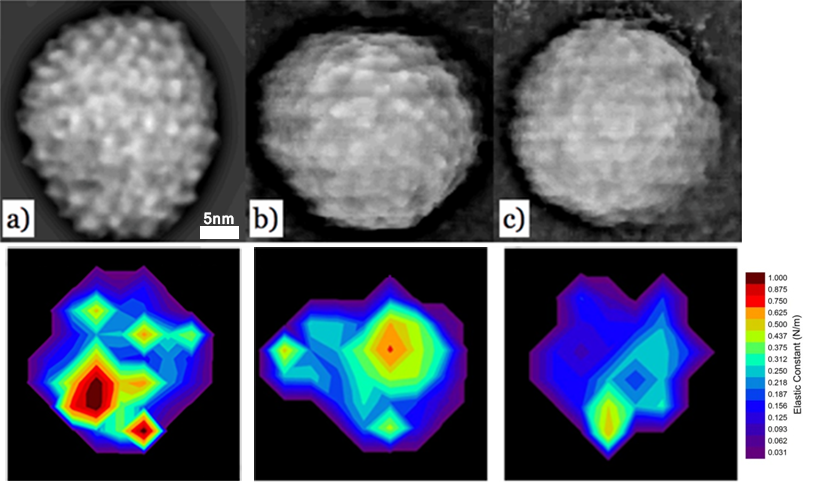
Subnanometer resolution AFM images reveal individual surface spikes formed by 4-helix bundles comprised of dimers of alpha-helical coiled coils of the core proteins on Hepatitis B virus (HBV) capsid (a) in solution. These distinctive features were observable in a smaller measure at Au-NP filled virus-like particles (VLPs) surfaces (b) and were virtually absent from RNA-filled VLP surfaces (c). Bottom images show elasticity mapping on single particles of HBV capsid, Au-NP filled VLP and RNA-filled VLP.
Elasticity mapping on single particles reveals mechanistic details of hepatitis B virus and virus-like particles
Ma, X., Li, H., Zlotnick, A. & Dragnea, B.
0000
(2016)
Ma, X., Li, H., Zlotnick, A. & Dragnea, B.
in prep
00
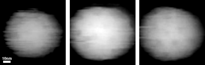
In situ AFM images show surface spike arrangement (dimples) of Sindbis virus (SINV) (a), and SINV conjugated with fluorescent proteins: Apple (b) and Venus (c).
Fusion of mApple and Venus fluorescent proteins to the Sindbis virus E2 protein leads to different cell-binding properties
Tsvetkova, I., Cheng, F., Ma, X., Moore, A., Howard, B., Mukhopadhyay, S., & Dragnea, B.
138-146
(2013)
Tsvetkova, I., Cheng, F., Ma, X., Moore, A., Howard, B., Mukhopadhyay, S., & Dragnea, B.
Virus Research
177