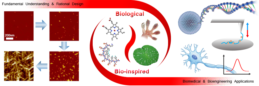
The Ma research group is interested in developing new classes of materials and nanostructures, with an emphasis on novel bio-inspired materials, for biomedical and bioengineering applications, such as biosensing, bioimaging, and drug delivery. We are also interested in understanding the fundamental issues of biomaterial self-assembly and structural growth that will enable the rational design of material sequence, micro/nano-structure, property and functionality. Our research is highly interdisciplinary, involving biochemistry, biophysics, biomaterials, biomedicine and bioengineering.
Our current research is focused on four major directions: (1) Fundamental studies of the self-assembly and mechanical properties of biological/bio-inspired materials for rational design; (2) Ultra-sensitive nano-scaled biosensors for the detection of disease biomarkers, and continuous in vivo monitoring of important biomolecules, such as glucose and neurotransmitters; (3) Bioimaging probes and drug delivery systems; (4) Investigation of the mechanisms of enzyme catalysis for drug development.
We approach these problems using a combination of modern scanning probe microscopy (in situ atomic force microscopy, electrostatic force microscopy, dynamic force spectroscopy, etc.), spectroscopic and electrochemical techniques to understand the structural, mechanical and electrochemical properties of biomaterials, as well as thermodynamic and kinetic controls on the self-assembly pathways, which will guide further development of bio-inspired materials and ultra-sensitive biosensors for biomedicine and nanotechnology.
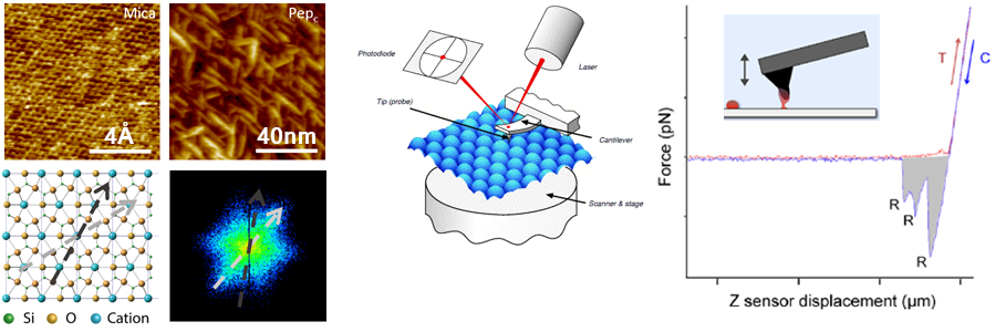
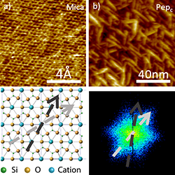
In this direction, we use atomic force microscopy (AFM) as a major instrument for fundamental studies of self-assembly pathways and mechanical properties of biological and bio-inspired materials. These fundamental studies will be used to facilitate rational design of bio-inspired materials for biomedical and bioengineering applications. AFM was first invented in 1983, but not until 2000, its application field reached biology and materials science, thanks to the novel design and development of the instrument, especially the "in situ" capability and extreme sensitivity in force measurements.
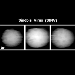
An area of particular interest to us is the self-assembly of biological and bio-inspired materials. Many essential biomolecular complexes and structures, such as ribozymes, proteasomes and viruses, involve a self-assembly process to form an organized structure. Understanding their self-assembly pathways is thus crucial for deciphering the molecular mechanisms underlying many important biological processes. Using in situ AFM, we directly observe the assembly and function of these molecular complexes in real time and at molecular level. These experiments allow us to examine structural transitions at the single-molecule level that are difficult to be detected by classical ensemble experiments, to probe the dynamic interactions between DNA, RNA and proteins, and to determine the relationship between the structural dynamics and function for these molecular complexes. This approach can also be applied to study self-assembly of bio-inspired materials, such as peptoids and AApeptides.
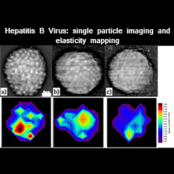
Besides imaging, AFM is well capable of measuring forces at pico-Newton (1pN = 10-12 N) and nano-Newton level, which falls into the range of most biomolecular interactions, including protein folding, protein-protein, protein-ligand and protein-nucleic acid interactions, cell adhesion, and virus elasticity. In this lab, we have studied the adhesive force development of bacterial holdfasts, and elastic behavior of brome mosaic virus (BMV) and hepatitis B virus (HBV).
AFM Image Gallery
Selected publications:
Ma, X., Zhang, S., Jiao, F., Newcomb, C., Zhang, Y., Prakash, A., Liao, Z., Baer, M., Mundy, C., Pfaendtner, J., Noy, A., Chen, C., & De Yoreo, J.
Berne, C., Ma, X., Licata, N.A., Neves, B., Setayeshgar, S., Brun, Y.V., & Dragnea, B.
Ni, P., Wang, Z., Ma, X., Das, N., Sokol, P., Chiu, W., Dragnea, B., Hagan, M., & Kao, C.C.
Vaughan, R., Tragesser, B., Ni, P., Ma, X., Dragnea, B., & Kao, C.C.
Tsvetkova, I., Cheng, F., Ma, X., Moore, A., Howard, B., Mukhopadhyay, S., & Dragnea, B.
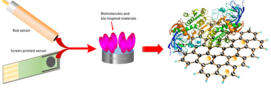
Biosensors are analytical devices where a biological/bio-inspired material is integrated within a physicochemical transducer, which may be optical, electrochemical, thermometric, piezoelectric, magnetic or micromechanical. Biosensors are used for the detection of an analyte, by yielding a digital electronic signal which is proportional to the concentration of the analyte. Examples of biosensors include immunosensors, glucose biosensors, DNA biosensors, microbial biosensors, and biosensors in food analysis.
A great challenge in this field is to fabricate fast, efficient, effective, low-cost and miniaturized biosensors for the in vivo detection and monitoring. In our lab, we focus on electrochemical and micromechanical biosensors. We develop novel bio-inspired materials, such as functionalized electro-conductive peptoids, graphene-like two-dimensional nanosheets and carbon/silicon-based nanohoops, to fabricate nano-scaled biosensors to detect disease biomarkers and continuously monitor important biomolecules, such as glucose and neurotransmitters, in vivo.
Selected publications:
Ma, X., Chen, T., Liu, L., & Li, G.
Liu, X., Xu, Y., Ma, X., & Li, G.
Ma, X., Liu, X., Xiao, H., & Li, G.
We are interested in synthesizing novel bioimaging probes, such as magnetic nanoparticles for magnetic resonance imaging (MRI), for cancer research. We are also interested in developing new drug delivery systems based on virus-like particles (VLPs).
Selected publications:
Our research in this direction focuses on the mechanisms and kinetics of catalytic reactions. We use a combination of in situ AFM and electrochemical techniques to investigate the energestic, structural and electrochemical properties of catalysts (e.g., enzymes). Understanding detailed mechanisms of catalytic reactions can facilitate rational design of efficient and robust catalysts for applications in the fields of electronic devices, energy production and storage, sensing and biomedicine.
Selected publications: